Knocheninfektion & gestufte Rekonstruktion
Ein stufenweises Protokoll, das kulturgeführten antibiotischen Knochenzement mit der induzierten Membrantechnik kombiniert – es erreicht lokale Antibiotikaspiegel, die eine systemische Therapie nicht erzielen kann, und baut dann das wieder auf, was die Infektion zerstört hat.
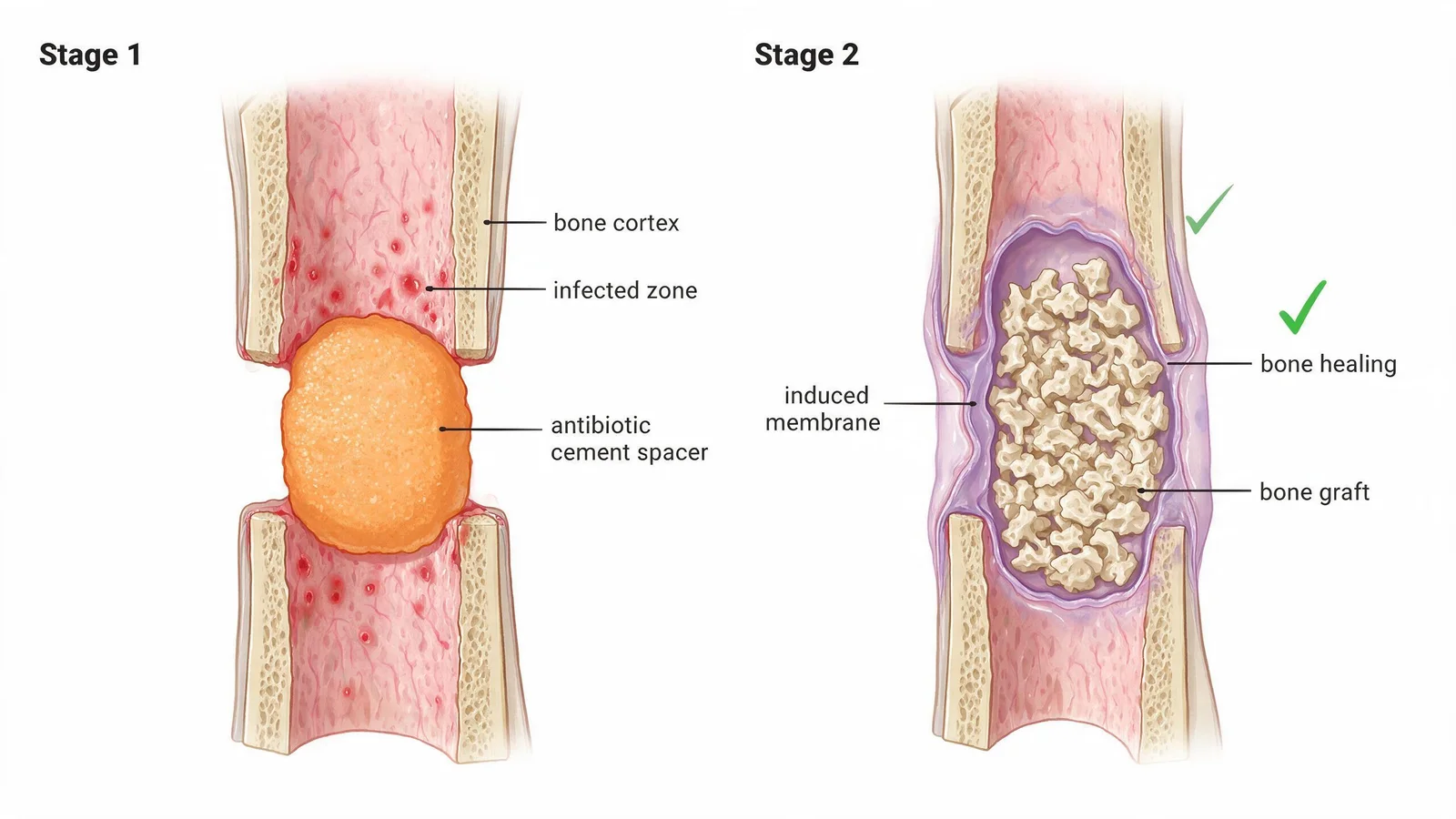
Die diabetische Fußosteomyelitis (DFO) ist eine der schwierigsten Wunden in der Medizin. Sie kombiniert Knochenzerstörung, bakteriellen Biofilm, schlechte lokale Durchblutung und beeinträchtigte Immunantwort. Standard-IV-Antibiotika erreichen oft Konzentrationen weit unterhalb der minimalen Hemmkonzentration im infizierten Knochen – insbesondere im diabetischen Fuß, wo die Durchblutung bereits reduziert ist. Unser stufenweises Protokoll geht dies direkt an.
Stufe 1 – Chirurgisches Debridement und Antibiotika-Spacer
Alles nicht lebensfähige Knochen- und Weichteilgewebe wird chirurgisch entfernt. Der Defekt wird mit einem antibiotikabeladenen Knochenzement-Spacer gefüllt – einer Perle oder einem Block, der mit kulturgeführten Antibiotika formuliert ist (typischerweise Vancomycin ± Tobramycin, angepasst an die Erregerempfindlichkeit). Der Spacer gibt über 4–8 Wochen hohe lokale Medikamentenkonzentrationen ab, während das umgebende Gewebe eine biologische induzierte Membran bildet.
- Knochenbiopsie beim Debridement zur Identifizierung von Erregern und Empfindlichkeiten (nicht nur Wundabstrich)
- Zementspacer innerhalb von 24 Stunden nach Kulturergebnis formuliert – abgestimmt auf den tatsächlichen Erreger
- Lokale Konzentrationen >2× MHK am Infektionsort aufrechterhalten
- Systemische Antibiotika parallel für 4–6 Wochen fortgesetzt
Stufe 2 – Rekonstruktion mit induzierter Membran
Nach 6–8 Wochen, sobald sich die Infektionsmarker (CRP, BSG, Leukozyten) normalisiert haben, wird der Zementspacer entfernt. Die induzierte Membran, die sich um ihn gebildet hat, sondert Wachstumsfaktoren ab, die das Transplantat vaskularisieren. Spongiosatransplantat (autolog oder synthetisch) wird eingebracht und die Membran wird verschlossen genäht – es entsteht ein biologischer Reaktor, der die Knochenregeneration beschleunigt.
Weichteilbedeckung
Nach der Knochenrekonstruktion wird jeder verbleibende Weichteildefekt mit lokalen Lappen, Spalthauttransplantation oder epidermaler Zellgewinnung je nach Defektgröße verschlossen. Ziel ist eine dauerhafte, belastungsfähige Bedeckung, die den rekonstruierten Knochen schützt.
Die Masquelet-Technik – Verwendung eines antibiotikabeladenen Knochenzement-Spacers zur Induktion einer biologischen Membran, gefolgt vom Ersatz des Spacers durch Knochentransplantat in einem zweiten Schritt – wird im Westen bei traumatischem Knochenverlust praktiziert. Ihre systematische Anwendung bei diabetischer Fußosteomyelitis mit MDR-Erregern, kombiniert mit kulturgeführten Antibiotika-Perlenformulierungen, ist außerhalb spezialisierter DFU-Rekonstruktionszentren nicht Routine. Unser Team hat ein spezifisches Protokoll für die diabetische Knochenumgebung entwickelt, in der Vaskularität und Weichteilintegrität zusätzliche Einschränkungen darstellen.
References
- Masquelet AC. Induced membranes for bone defect reconstruction. J Orthop Surg Res. 2019;14:405.
- Lew DP, Waldvogel FA. Osteomyelitis. Lancet. 2004;364(9431):369-379.
- Lipsky BA et al. IWGDF guidance on the diagnosis and management of bone infection (osteomyelitis) in persons with diabetes. Diabetes Metab Res Rev. 2020;36(S1):e3282.
Was kostet das?
Die Gesamtkosten an Chinas spezialisierten DFU-Zentren – einschließlich stationärem Aufenthalt, Eingriffen und Koordination – sind in der Regel weitaus niedriger als vergleichbare private Behandlungen in den USA, Großbritannien oder der EU. Die meisten Patienten zahlen selbst; wir unterstützen auch bei der Dokumentation für die Kostenerstattung durch die Versicherung. Wir stellen Ihnen eine transparente, detaillierte Kostenschätzung zur Verfügung, bevor Sie sich zu etwas verpflichten – kostenlos.
Kostenlose Kostenschätzung anfordern →Kostenlose Fallprüfung anfordern
Unser Koordinationsteam wird Ihren Fall prüfen und innerhalb von 24–48 Stunden antworten.
Diese Website bietet ausschließlich medizinische Informationen zur wissenschaftlichen Aufklärung. Sie bietet KEINE Online-Diagnosen, Rezepte oder medizinische Beratung. Konsultieren Sie einen approbierten Arzt für jede medizinische Entscheidung.