Bone Infection & Staged Reconstruction
A staged protocol combining culture-directed antibiotic bone cement with the induced-membrane technique — achieving local antibiotic levels that systemic therapy cannot reach, then rebuilding what infection destroyed.
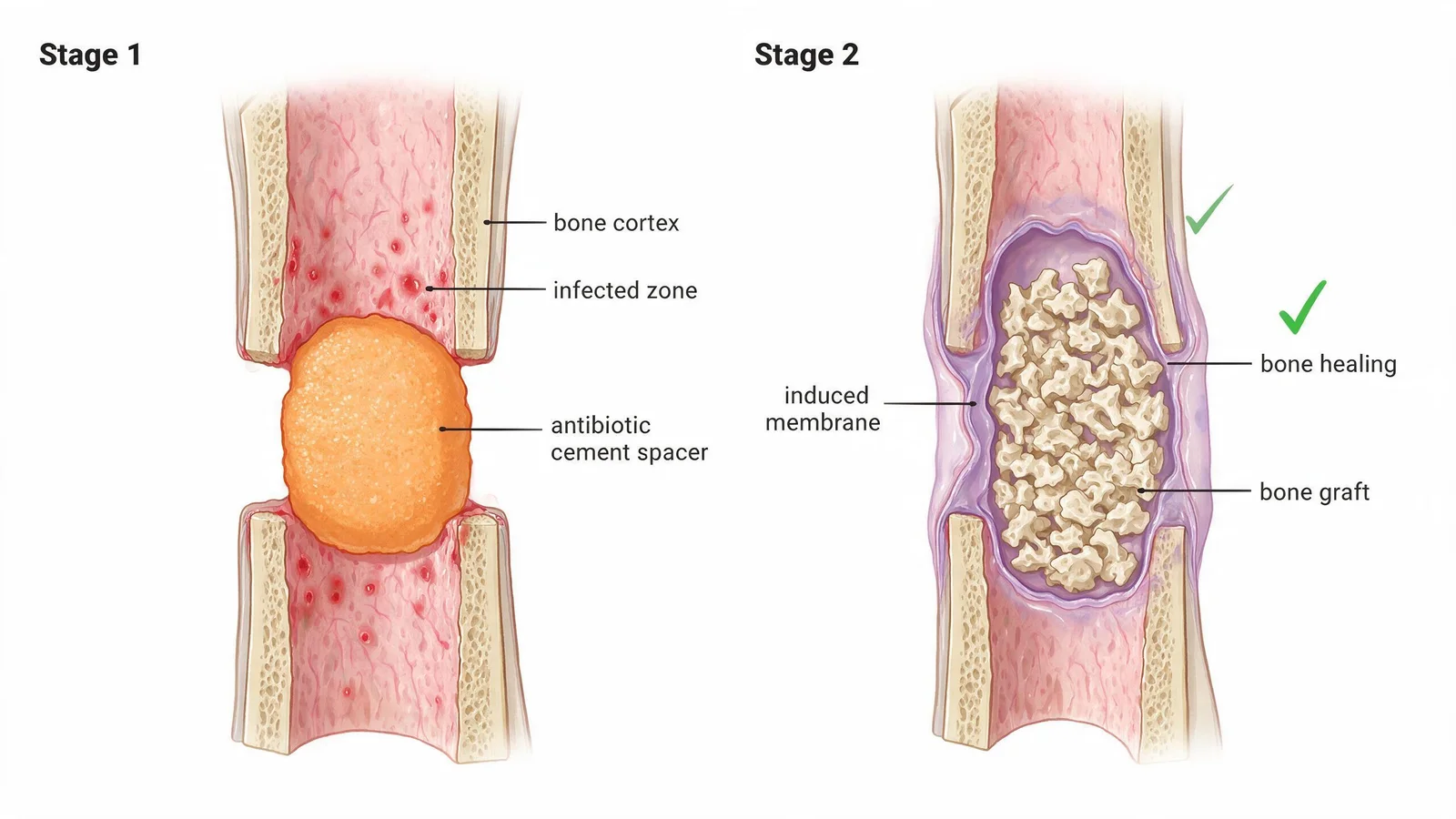
Diabetic-foot osteomyelitis (DFO) is one of the most difficult wounds in medicine. It combines bone destruction, bacterial biofilm, poor local perfusion, and compromised immune response. Standard IV antibiotics often achieve concentrations far below the minimum inhibitory concentration inside infected bone — particularly in the diabetic foot where perfusion is already reduced. Our staged protocol addresses this directly.
Stage 1 — Surgical debridement and antibiotic spacer
All non-viable bone and soft tissue is surgically removed. The defect is filled with an antibiotic-loaded bone cement spacer — a bead or block formulated with culture-directed antibiotics (typically vancomycin ± tobramycin, adjusted to organism sensitivity). The spacer releases high local drug concentrations over 4–8 weeks while the surrounding tissue forms a biological induced membrane.
- Bone biopsy taken at debridement to identify organism and sensitivities (not just wound swab)
- Cement spacer formulated within 24h of culture results — matched to the actual pathogen
- Local concentrations >2× MIC maintained at the infection site
- Systemic antibiotics continued in parallel for 4–6 weeks
Stage 2 — Induced membrane reconstruction
At 6–8 weeks, once infection markers (CRP, ESR, WBC) normalize, the cement spacer is removed. The induced membrane that has formed around it secretes growth factors that vascularize the graft. Cancellous bone graft (autologous or synthetic) is placed, and the membrane is sutured closed — creating a biological reactor that accelerates bone regeneration.
Soft-tissue coverage
After bone reconstruction, any remaining soft-tissue defect is closed with local flaps, split-skin grafting, or epidermal cell harvesting depending on defect size. The goal is durable, load-bearing coverage that protects the reconstructed bone.
The Masquelet technique — using an antibiotic-loaded bone cement spacer to induce a biological membrane, then replacing the spacer with bone graft in a second stage — is practiced in the West for traumatic bone loss. Its systematic application to diabetic-foot osteomyelitis with MDR organisms, combined with culture-directed antibiotic bead formulations, is not routine outside dedicated DFU reconstruction centers. Our team has developed a specific protocol for the diabetic bone environment, where vascularity and soft-tissue integrity are additional constraints.
References
- Masquelet AC. Induced membranes for bone defect reconstruction. J Orthop Surg Res. 2019;14:405.
- Lew DP, Waldvogel FA. Osteomyelitis. Lancet. 2004;364(9431):369-379.
- Lipsky BA et al. IWGDF guidance on the diagnosis and management of bone infection (osteomyelitis) in persons with diabetes. Diabetes Metab Res Rev. 2020;36(S1):e3282.
Doctors who perform this treatment

What does this cost?
All-in costs at China's specialist DFU centres — including inpatient stay, procedures, and coordination — are typically far lower than equivalent private care in the US, UK, or EU. Most patients self-pay; we also support insurance reimbursement documentation. We provide a transparent itemised estimate before you commit to anything, at no charge.
Request a free cost estimate →Free medical record review
Share your diabetic foot ulcer records for a free specialist review. Our care coordination team responds within 24–48 hours with an honest assessment.
This website provides medical-information science popularization only. It does NOT provide online diagnosis, prescriptions, or medical advice. Consult a licensed physician for any medical decision.