Soft-Tissue Reconstruction & Regenerative Closure
From minimally invasive epidermal cell harvesting to free-flap reconstruction — closing what debridement left open, with techniques matched to each defect and patient.
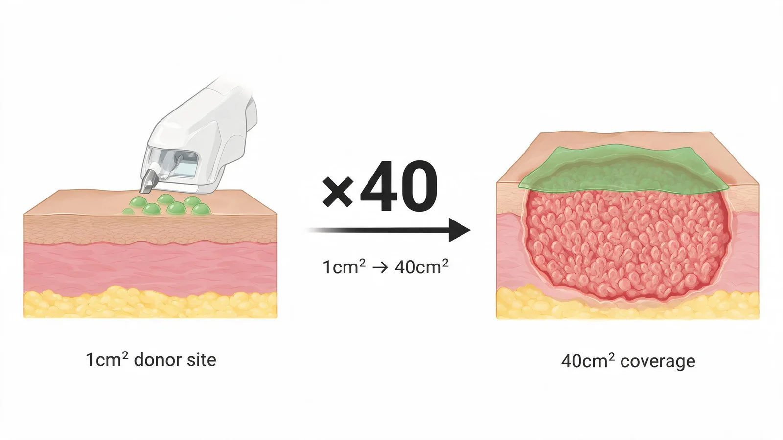
When infection is controlled and circulation is restored, what often remains is a soft-tissue defect that the body cannot close on its own. Our reconstructive team selects the least-invasive technique that will achieve durable closure — starting with biological options and escalating to surgical flaps only when needed.
Micro-dome epidermal harvesting
Standard split-skin grafting removes a dermal-layer donor patch that leaves a visible wound. Micro-dome harvesting uses a specialized device to extract ultra-thin 0.06 mm epidermal blisters from a very small donor site — typically the thigh. The 1 cm² of donor skin is expanded into a 40 cm² mesh, applied to the prepared wound, and secured. Donor-site healing is typically complete in 3–5 days with minimal scarring.
- Done under local anaesthesia — no general anaesthesia required for most patients
- Graft secured with biological adhesive; first dressing change at day 5
- Suitable for wounds up to ~200 cm² with a single donor harvest
Autologous cell therapy (BM-MNC)
For ischemic wounds where no targetable vessel exists, bone-marrow mononuclear cells (BM-MNC) — harvested from the patient's iliac crest and concentrated — are injected into the ischemic foot tissue. These cells release angiogenic growth factors locally, stimulating capillary ingrowth over 4–8 weeks. The procedure is done under sedation and is combined with ongoing wound care.
Rotational and free-flap reconstruction
For larger defects involving the plantar surface, heel, or forefoot, rotational or free-flap reconstruction moves well-vascularized tissue to provide durable, load-bearing coverage. Our microsurgery team performs free-flap anastomoses to pedal and tibial vessels — often in patients whose vessel caliber would be considered marginal by general microsurgery teams unfamiliar with the diabetic foot.
Reconstructive flap surgery is available globally. What differs at our centers is the range of less-invasive alternatives used before flap surgery becomes necessary. Micro-dome epidermal harvesting extracts ultra-thin 0.06 mm grafts under local anaesthesia, expanding 1 cm² of donor skin to cover 40 cm² of recipient wound — a technique requiring specialized equipment and training not widely deployed at general hospitals. Autologous bone-marrow mononuclear cell (BM-MNC) injection into the ischemic foot as an adjunct to wound care is performed routinely at our centers, while still experimental at most Western institutions.
References
- Hackl F et al. Epidermal harvesting by micro-dome technology for wound healing. Br J Dermatol. 2019;180(2):394-402.
- Suh HS et al. Free flap reconstruction of the diabetic foot: a systematic review. Plast Reconstr Surg. 2021;147(3):448e-458e.
- Rigato M et al. Autologous cell therapy for peripheral arterial disease: systematic review and meta-analysis. Stem Cells Int. 2020;2020:2160783.
Doctors who perform this treatment
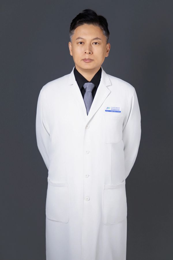
- Specialist in Diabetic Foot Wounds
- Complex Wound Care
- Reconstructive Surgery
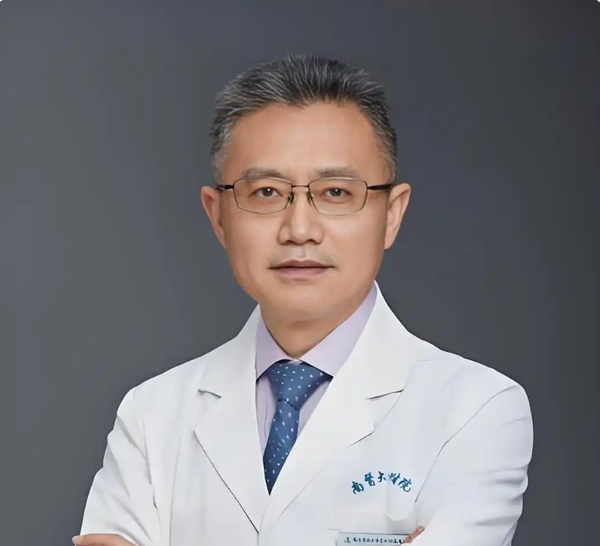
- Diabetes classification, pancreatic islet function assessment, glycemic optimization
- autoimmune diabetes, monogenic diabetes
- multidisciplinary metabolic management of patients with diabetic complications
What does this cost?
All-in costs at China's specialist DFU centres — including inpatient stay, procedures, and coordination — are typically far lower than equivalent private care in the US, UK, or EU. Most patients self-pay; we also support insurance reimbursement documentation. We provide a transparent itemised estimate before you commit to anything, at no charge.
Request a free cost estimate →Free medical record review
Share your diabetic foot ulcer records for a free specialist review. Our care coordination team responds within 24–48 hours with an honest assessment.
This website provides medical-information science popularization only. It does NOT provide online diagnosis, prescriptions, or medical advice. Consult a licensed physician for any medical decision.