إعادة بناء الأنسجة الرخوة والإغلاق التجديدي
من حصاد الخلايا البشروية بالتدخل الجراحي البسيط إلى إعادة البناء بالطعوم الحرة — إغلاق ما يتركه التنضير مفتوحًا، بتقنيات تتناسب مع كل عيب ومريض.
عندما يتم السيطرة على العدوى واستعادة الدورة الدموية، غالبًا ما يتبقى عيب في الأنسجة الرخوة لا يستطيع الجسم إغلاقه من تلقاء نفسه. يختار فريقنا الترميمي التقنية الأقل تدخلاً التي تحقق إغلاقًا دائمًا — بدءًا بالخيارات البيولوجية والتصعيد إلى الطعوم الجراحية فقط عند الحاجة.
الحصاد الجلدي البشروي الدقيق
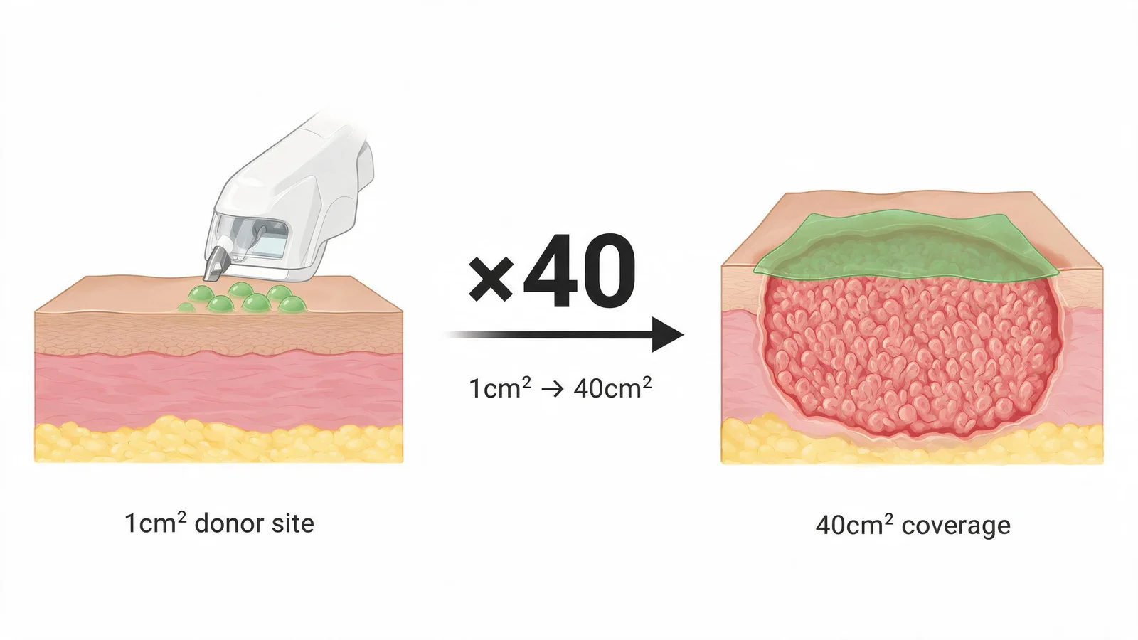
يزيل الطعم الجلدي المنقسم القياسي رقعة من الطبقة الأدمية تترك جرحًا ظاهرًا. يستخدم الحصاد الدقيق جهازًا متخصصًا لاستخراج بثور بشروية رقيقة جدًا بسمك 0.06 مم من موقع مانح صغير جدًا — عادةً الفخذ. يتم توسيع 1 سم² من الجلد المانح إلى شبكة بمساحة 40 سم²، وتوضع على الجرح المُجهز وتُثبت. عادةً ما يكتمل التئام موقع المانح في 3-5 أيام مع ندبة ضئيلة.
- يتم تحت التخدير الموضعي — لا حاجة لتخدير عام لمعظم المرضى
- يُثبت الطعم بمادة لاصقة بيولوجية؛ أول تغيير للضمادة في اليوم الخامس
- مناسب للجروح حتى ~200 سم² مع حصاد واحد من المانح
العلاج الخلوي الذاتي (BM-MNC)
بالنسبة للجروح الإقفارية التي لا يوجد فيها وعاء مستهدف، يتم حقن الخلايا أحادية النواة من نخاع العظم (BM-MNC) — المستخرجة من عرف الحرقفة للمريض وتركيزها — في أنسجة القدم الإقفارية. تطلق هذه الخلايا عوامل نمو وعائية محليًا، مما يحفز نمو الشعيرات الدموية على مدى 4-8 أسابيع. يتم الإجراء تحت التخدير ويُدمج مع العناية المستمرة بالجروح.
إعادة البناء بالطعوم الدورانية والحرة
بالنسبة للعيوب الأكبر التي تشمل السطح الأخمصي أو الكعب أو مقدمة القدم، تنقل إعادة البناء بالطعوم الدورانية أو الحرة أنسجة جيدة الأوعية الدموية لتوفير تغطية دائمة تتحمل الوزن. يقوم فريق الجراحة المجهرية لدينا بإجراء مفاغرات للطعوم الحرة إلى الأوعية الأخمصية والظنبوبية — غالبًا في مرضى قد يعتبر قطر أوعيتهم هامشيًا من قبل فرق الجراحة المجهرية العامة غير المألوفة بالقدم السكري.
جراحة الطعوم الترميمية متاحة عالميًا. ما يختلف في مراكزنا هو نطاق البدائل الأقل تدخلاً المستخدمة قبل أن تصبح جراحة الطعوم ضرورية. الحصاد الجلدي البشروي الدقيق يستخرج طعومًا رقيقة جدًا بسمك 0.06 مم تحت التخدير الموضعي، مما يوسع 1 سم² من الجلد المانح لتغطية 40 سم² من الجرح المتلقي — وهي تقنية تتطلب معدات وتدريبًا متخصصًا غير منتشر على نطاق واسع في المستشفيات العامة. حقن الخلايا أحادية النواة الذاتية من نخاع العظم (BM-MNC) في القدم الإقفارية كإضافة إلى العناية بالجروح يتم بشكل روتيني في مراكزنا، بينما لا يزال تجريبيًا في معظم المؤسسات الغربية.
References
- Hackl F et al. Epidermal harvesting by micro-dome technology for wound healing. Br J Dermatol. 2019;180(2):394-402.
- Suh HS et al. Free flap reconstruction of the diabetic foot: a systematic review. Plast Reconstr Surg. 2021;147(3):448e-458e.
- Rigato M et al. Autologous cell therapy for peripheral arterial disease: systematic review and meta-analysis. Stem Cells Int. 2020;2020:2160783.
ما هي التكلفة؟
التكاليف الإجمالية في مراكز قدم السكري المتخصصة في الصين — بما في ذلك الإقامة والإجراءات والتنسيق — أقل عادةً بكثير من الرعاية الخاصة المماثلة في الولايات المتحدة أو المملكة المتحدة أو الاتحاد الأوروبي. يتكفل معظم المرضى بالتكاليف بأنفسهم، كما ندعم توثيق استرداد التأمين. نقدم تقديراً شفافاً مفصّلاً قبل الالتزام بأي شيء، دون أي رسوم.
طلب تقدير تكلفة مجاني ←طلب مراجعة مجانية للحالة
سيراجع فريق التنسيق الطبي لدينا حالتك ويرد في غضون 24–48 ساعة.
يقدّم هذا الموقع معلومات طبية تثقيفية فحسب، ولا يُقدّم تشخيصاً عبر الإنترنت أو وصفات طبية أو مشورة علاجية. يُرجى استشارة طبيب مرخّص لاتخاذ أي قرار طبي.